
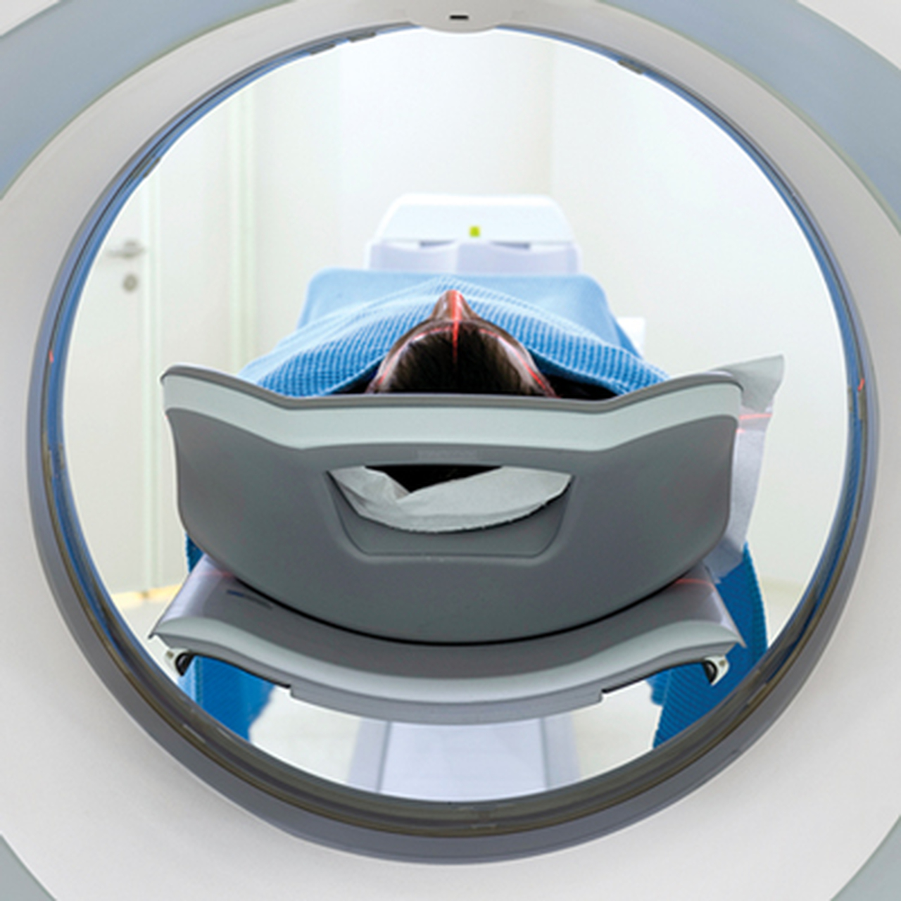
Description
+ Include: 86 videos + 1 pdf, size: 42.1 GB
+ Target Audience: radiologists
+ Sample video: contact me for sample video
+ Information:
Stay Ahead in Your Field
NYU’s Head to Toe Imaging is extremely thorough and will help you keep up with recent developments and current trends in the field. Led by H. Quinny Cheng, MD, this CME program focuses on the practical aspects of established CT, MR, US, PET, and interventional techniques, as well as new techniques and applications for these modalities. It will help you to better:
Evaluate and fit current imaging techniques and protocols for sub specialty imaging into clinical practice
Explain the Bosniak classification system for small renal masses
Develop imaging protocols to optimize radiation safety and dose reduction in CT
Utilize MR imaging in apt patient populations without compromising diagnostic accuracy
Strategize to optimize and incorporate a gamut of studies, including CT, MR and PET to accurately stage neoplasms
Expand Your Skills
Date of Original Release: February 15, 2020
+ Topics:
1. Pediatric Brain Tumors – What is New.mp4
2. Adult Brain Tumors – Radiogenomics.mp4
3. Stroke Imaging.mp4
4. Adult Acquired White Matter Disorders.mp4
5. Metastatic Neck Disease – Assessment of Unknown Primary.mp4
6. Sinuses – Inflammatory Diseases & Post Operative Imaging.mp4
7. Transverse Myelitis.mp4
8. AJCC Update in HN Cancer Staging.mp4
9. Post-operative Surveillance in HN Tumors.mp4
10. Prostate Artery Embolization – From Diagnostic Workup to Post-Procedure Imaging.mp4
11. Imaging & Management of Portal Hypertension.mp4
12. LI-RADS & Interventional Oncology Follow-Up.mp4
13. Challenging IR Cases.mp4
14. Neuroradiology ED Cases.mp4
15. Non-Vascular – Brain & Spine Cases.mp4
16. Vascular – Brain & Spine Cases.mp4
17. Head & Neck Cases.mp4
18. Imaging of the Postoperative Breast.mp4
19. Tips & Tricks for Sonographic Correlation.mp4
20. Mammographic Analysis of Breast Calcifications.mp4
21. Diagnostic Evaluation with Tomosynthesis.mp4
22. Mammographic Follow-Up as an Alternative to Biopsy – Strengths, Limitations & Pitfalls.mp4
23. Hormones, Imaging Biomarkers & Breast Cancer Risk.mp4
24. A New Perspective on Male Breast Cancer.mp4
25. Updates on Fast Breast MRI & Advanced Breast MRI Techniques.mp4
26. Medicolegal Aspects of Breast Imaging.mp4
27. Pediatric Hip.mp4
28. Pediatric Knee.mp4
29. Pediatric Elbow.mp4
30. Pediatric Cases.mp4
31. Breast Imaging of the Young, Pregnant & Lactating Patient.mp4
32. Challenging Image-guided Breast Procedure Cases.mp4
33. Shoulder Imaging – Pearls & Pitfalls.mp4
34. Elbow Imaging – Pearls & Pitfalls.mp4
35. Groin Injuries.mp4
36. Hip Cartilage & Labral Injuries – What the Surgeon Wants to Know.mp4
37. Knee Avulsion Injuries.mp4
38. AI in MSK Imaging.mp4
39. MSK Infections – How to Avoid Misdiagnosis.mp4
40. Surgery of the Lumbar Spine – The Basics.mp4
41. Weird Bone Lesions.mp4
42. Ultrasound of Lumps & Bumps.mp4
43. Ultrasound Guided Procedures.mp4
44. PET Imaging of Colorectal Carcinoma.mp4
45. PET Imaging of Prostate Cancer.mp4
46. Management of Neuroendocrine Tumors with Ga-Dotatate PET.mp4
47. MSK Cases – Upper Extremity.mp4
48. PET Results & Path Reports – Concordance or Learning Opportunity.mp4
49. MSK Cases – Lower Extremity.mp4
50. Ga-Dotatate PET – False-positives & Incidental Malignancies.mp4
51. Central Airway Diseases.mp4
52. An Imaging Approach to Small Airway Disease.mp4
53. Seeing the Picture in a Mosaic.mp4
54. Thoracic Sarcoidosis – Hidden in Plain View.mp4
55. Shades of Gray – Subsolid Lung Nodules.mp4
56. Leveling the Playing Field – Navigation Guided Thoracic Intervention.mp4
57. State of the Art – CT Pulmonary Angiography.mp4
58. Smoke – Differentiating Flow Artifact from Pathology on Thoracic CTA.mp4
59. Coronary Anomaly Misses & Misperceptions.mp4
60. Challenging Cardiac CT Cases.mp4
61. Extraordinary Cases in Congenital Heart Disease.mp4
62. Challenging Chest Cases.mp4
63. Segmental Approach to Congenital Heart Disease.mp4
64. Zebras in the Chest Reading Room.mp4
65. Cardiovascular MRI Cases – What We Got Wrong & Why.mp4
66. Practicing Green CT.mp4
67. Ultrasound Diagnosis of Non-gynecologic Causes of Pelvic Pain.mp4
68. Small Bowel Imaging.mp4
69. Imaging of Solid Renal Masses – Lessons Learned from Bosniak Classification.mp4
70. Prostate MR.mp4
71. Bariatric Imaging Pearls from the Practicing Surgeon.mp4
72. Top 10 Updates in Body Imaging from 2019.mp4
73. Challenging Benign Focal Liver Lesions.mp4
74. Fibrosis, Cirrhosis & HCC – An Imaging Update.mp4
75. Post Y90 Liver Imaging.mp4
76. Rectal MRI.mp4
77. Mass Casualty Incidents – An Introduction for Imagers.mp4
78. Facial Trauma.mp4
79. Emergency Pelvic US.mp4
80. Weird & Wonderful ER Cases.mp4
81. TI-RADS.mp4
82. Renal Cases.mp4
83. Vascular Cases.mp4
84. Bowel Cases.mp4
85. Gynecologic Cases.mp4
86. Body ER Cases.mp4
Syllabus.pdf




Reviews
There are no reviews yet.